Venous duplex ultrasound is an important tool when a clinician needs to understand how blood moves through the veins of the legs. The name sounds technical, but the question it addresses is practical: what is happening, and how might that change the treatment plan? When assessing non-surgical varicose vein treatment, a photograph of the skin cannot replace information about the circulation underneath it.
This guide explains what can be examined, how the appointment commonly works and what to ask when you receive the report. The scan is adapted to the clinical question. An examination requested for varicose veins does not have exactly the same purpose as one performed because a blood clot is suspected. Understanding why it was requested makes the result easier to interpret without treating every unfamiliar word as a cause for alarm.
Ultrasound and Doppler provide complementary information
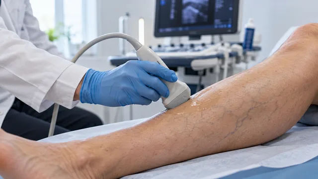
Ultrasound uses sound waves to show structures inside the body. The Doppler component supplies information related to blood movement. Together, they allow the professional to examine both anatomy and flow. Ionising radiation is not used, and the probe moves over the skin with gel to improve contact. The person conducting the examination adjusts the technique to the area and clinical question.
The colours on the screen do not simply represent “good” or “bad” blood, and colour alone does not identify an artery or a vein. They are part of a flow display that must be interpreted in relation to equipment settings and the examination. A striking sound does not necessarily indicate a serious abnormality either. What matters is the clinical interpretation of the overall study, rather than the impression created by a few seconds of screen activity.
What the scan may look for in varicose veins
A venous assessment can identify the segments involved and their connections. The examiner may investigate whether reflux is present, where it begins, its route and its relationship with the visible veins. The deep circulation is also assessed where relevant. This map can help determine whether a superficial trunk, particular branches or no structure at that time should be treated.
The information helps avoid choosing a procedure solely from the apparent size of a vein. Two legs that look similar can have different flow patterns. Likewise, a visible vein after earlier treatment may have a different origin from the original problem. In that situation, ultrasound helps formulate specific questions before deciding whether another intervention is justified, which technique might suit it and what benefit could reasonably be expected.
How the examination commonly takes place
Depending on the aim, parts of the scan may be performed lying down, sitting or standing. Position matters because it changes pressure and the behaviour of venous return. You may be asked to change position, or the examiner may use gentle manoeuvres to observe the response of blood flow. Mention difficulty standing, dizziness or mobility limitations so that the examination can be adapted safely.
Probe pressure may be uncomfortable over a tender area, but you should report pain rather than silently tolerate substantial discomfort. The appointment length depends on the question, whether one or both legs are examined and the complexity of the venous anatomy. Duration alone does not indicate quality or severity. A longer examination may simply reflect the need to document several different segments or to compare current findings with previous treatment.
Preparation: comfortable clothing and useful records
For a routine leg vein scan, clothing that makes the area easy to access is practical. Check the centre's instructions because preparation can differ when another investigation is performed at the same appointment. Do not assume that fasting is required, and do not stop medication unless specifically advised. A vascular examination of a different part of the body may have different preparation requirements.
Bring previous reports, particularly after thrombosis, surgery, sclerotherapy or another endovenous procedure. Dates and details of the technique provide valuable context. If you only have images without a written report, bring them and explain what you remember. The radiology and ultrasound service and the referring specialist should understand the reason for the scan so that the examination answers a useful clinical question instead of producing an isolated description.
Understanding the report without self-diagnosing
Terms such as “patent”, “reflux” and “saphenous” describe anatomical or functional findings. A patent vein allows blood to pass; reflux refers to backward flow under the conditions examined. A finding does not automatically require treatment. Its significance depends on symptoms, examination, context and the reasonable alternatives available for the person being assessed.
Ask which finding is the main one and which are secondary. Does the result explain the symptom that brought you to the appointment, and does it change the proposed plan? A normal result in the territory examined may be reassuring, but it does not answer every possible cause of pain or swelling. If symptoms persist, other explanations may need consideration. Understanding the limits of the report is as useful as understanding its technical terminology.
Limits of the scan and follow-up
Duplex ultrasound depends on a clear clinical question and appropriate performance and interpretation. It is not a universal picture of all aspects of vascular health. In selected situations, other investigations may be useful, but that does not mean everyone needs more tests. Each investigation should be justified by the information it is expected to provide and by whether that information could change management.
After some procedures, ultrasound is used to assess particular aspects of recovery. Review timing is individual. New acute symptoms should not be ignored because an earlier scan was reassuring: the report describes a particular territory at a particular point in time. Clinical changes remain important between examinations, and a new concern may require a different assessment from the one originally requested for routine varicose veins.
Understanding what happens inside the veins
Blood reaches the legs through the arteries and returns towards the heart through superficial and deep veins. Contractions of the calf muscles help move it upwards, while small valves encourage flow in the right direction. When some valves no longer close effectively, blood can flow backwards through a particular segment. This is called venous reflux and can contribute to increased pressure within parts of the venous network.
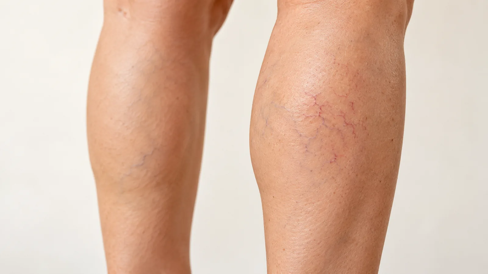
The vein visible at the surface does not necessarily explain the whole problem. It may be a branch connected to another vein with reflux, or a small surface change without significant disease in the main superficial trunks. Counting visible veins or looking at a photograph therefore cannot establish the right treatment. An examination connects the appearance with symptoms, distribution and, where appropriate, ultrasound findings.
It is also useful to separate cosmetic concerns from functional effects. Someone may be troubled by small visible veins despite having very little discomfort. Another person may experience heaviness or swelling even though changes at the surface look relatively modest. Both deserve a clear explanation, but they do not automatically require the same investigation, priority or intervention. Symptom intensity is not a precise measurement of the risk of complications either.
Family history, pregnancy, age, previous vascular events and long periods of immobility may all contribute to the overall picture. They do not establish a single cause, and they do not mean that the person has done something wrong. The consultation identifies which factors matter in that individual situation and which can realistically be changed. Understanding this distinction makes it easier to see why everyday measures may ease discomfort without removing an established vein, while a procedure targets a particular part of the circulation. Neither approach should be judged only by how a leg looks in one photograph.
What a complete vascular assessment contributes
An assessment begins with how the problem started, what changes the symptoms and how it affects everyday life. Useful details include whether one leg or both are affected, whether discomfort builds towards the evening, whether walking helps and whether anything has changed recently. Previous blood clots, procedures, pregnancies, medicines and other health conditions are also relevant to circulation and to the safety of possible treatment.
The examination may include visible veins, swelling, skin colour, temperature and skin condition. The clinician may check pulses when this helps assess arterial circulation. Leg discomfort is not always caused by veins: muscle, joint, nerve and lymphatic problems can produce overlapping symptoms. Several causes may exist together, in which case a particularly clear discussion about expectations is important before choosing an intervention.
Duplex ultrasound combines images of the structures with information about blood flow. When indicated, it can assess whether veins are open, the direction of flow and possible sites of reflux. Its purpose is not simply to add a routine test. It should answer questions that could change the diagnosis or the choice of treatment. The findings must be interpreted alongside the history and examination, rather than as a collection of isolated measurements.
Before making a decision, ask for an understandable explanation: which vein is involved, whether the symptoms fit that finding and what treatment is expected to achieve. It is equally reasonable to ask which symptoms might remain if they have a different cause. A sound decision includes alternatives, sometimes including observation. The appointment should end with a practical plan, a reason for each next step and clear advice about when to return. Knowing what uncertainty remains is useful information, not a sign that the consultation has failed to provide an answer.
Comparing conservative care and procedures
Conservative measures and procedures address different goals. Suitable activity, position changes, skin care and prescribed compression may help manage discomfort. They do not necessarily correct the reflux responsible for the problem or remove an established varicose vein. Where intervention is appropriate, the choice depends on anatomy, symptoms, medical history and the person's informed preferences, rather than on a procedure name alone.
Options used for venous disease include thermal endovenous techniques, sclerotherapy and selected surgical approaches. No single method suits every vein. Its location, course, size, relationship to skin and nerves, and the condition of the deep circulation can all change the recommendation. The fact that a technique is available at a clinic does not mean that every patient attending that clinic should receive it.
Useful comparisons cover the main objective, anaesthesia, expected number of visits, likely discomfort, temporary restrictions and follow-up. When a price is provided after assessment, it should explain what is included: investigations, the procedure, materials, reviews and any potential additional treatment. A quotation does not replace a clinical indication, and it should not be interpreted as a promise of a complete or permanent result.
Possible unwanted effects should also be discussed. Depending on the procedure, these may include bruising, inflammation, pigmentation, local discomfort or altered sensation. Less common complications can require medical attention. The clinician should explain the risks relevant to the specific technique and individual circumstances rather than provide an identical list for every treatment. Asking about alternatives, including not intervening, helps put the expected benefit in perspective. Informed consent is an individual conversation as well as a document. Having time to understand the explanation and ask questions is part of that process, even when the proposed treatment is performed as an outpatient procedure.
Building a routine you can maintain
Small, repeatable changes are often more useful than an ambitious routine that cannot fit everyday life. Alternating sitting with movement, walking within personal ability and avoiding long uninterrupted periods of stillness can help leg comfort. Where mobility is limited, the plan should accommodate that limitation instead of imposing a universal step count or a fixed exercise target on everyone.
Ankle movement and calf muscle activity contribute to the muscle pump that supports venous return. Brief movement breaks can be incorporated into a working day when appropriate for the person's clinical situation. Every break does not need to become an intense workout. Gradual, tolerable activity is usually easier to sustain than occasional strenuous efforts followed by prolonged inactivity, and the routine should remain practical when work or family responsibilities change.
When compression is prescribed, the indication and fit matter. Greater pressure does not automatically mean greater benefit. Size, skin condition, the ability to put the garment on and possible arterial disease all need consideration. A garment causing substantial pain, marked colour change or skin injury should not simply be endured without advice. Copying a relative's prescription or extending a post-procedure regimen independently is not a reliable way to choose suitable compression.
Daily care also means noticing the condition of the skin and paying attention to new changes. Rest and comfortable leg elevation may ease some symptoms, but they do not replace assessment when swelling persists or increases. These measures support a clinical plan. They do not guarantee that new varicose veins will never develop, and they do not mean that every symptom should be managed through lifestyle changes alone. A useful routine is one that supports function, can be maintained and has clear limits about when further professional advice is needed.
Warning signs that change the urgency
Not every leg problem should wait for a routine appointment. New swelling that is clearly greater in one leg, especially with pain, warmth or a change in colour, needs prompt medical assessment. Having known varicose veins does not make it safe to attribute every new symptom to them. A photograph or a messaging conversation cannot rule out a deep vein thrombosis either.
Breathlessness, chest pain, fainting or coughing up blood require urgent medical help. In Spain, the emergency number is 112. Trying to manage these symptoms by walking, massaging the leg or taking someone else's medication is not appropriate. An acute problem needs assessment on its own merits, even when a person already has a familiar explanation for occasional leg discomfort.
Bleeding from a varicose vein also calls for action. Lie down, raise the leg if possible and apply firm direct pressure with clean material over the bleeding point. Urgent help is needed if bleeding does not stop, is substantial or the person feels unwell. Even if it settles, the cause should be reviewed to reduce the risk of another episode. A homemade tourniquet should not be used.
Other changes can justify urgent or priority assessment: a wound that is not healing, a red and hot area accompanied by fever or increasing pain, or a foot that suddenly becomes cold, pale and painful. These examples explain the limits of general information; they are not a checklist for making a diagnosis at home. If the seriousness is uncertain, contact a healthcare service able to advise on urgency. A clinic's ordinary appointment channel is not a substitute for emergency services, and waiting for a reply to a non-urgent form is not appropriate when potentially serious symptoms are developing.
Assessing progress without promises of instant results
Follow-up should compare changes that matter to the agreed goal. If the priority was less heaviness, useful questions concern how a working day feels, whether walking is easier and whether rest is more comfortable. If appearance was also a goal, photographs taken under similar conditions may help document change. None of these observations replaces examination when new symptoms develop or the pattern changes unexpectedly.
Some changes after a procedure cannot be interpreted reliably in the first few days. Swelling, bruising or areas of firmness may temporarily affect appearance. The treating team should explain the expected course for the actual technique used and which findings warrant contact before the planned review. It is unwise to assume that everything is normal because treatment was minimally invasive, just as a small bruise does not by itself demonstrate treatment failure.
Reviews may involve a clinical assessment and, where indicated, repeat ultrasound. Their timing should not be copied from someone else's experience because the procedure, medical history and findings all matter. Keeping the initial report, details of the technique and written instructions supports continuity of care. If another centre is consulted later, those documents can reduce incomplete interpretation and unnecessary duplication of investigations.
Venous disease can change over time. Treating one vein does not remove the underlying tendency to develop other abnormalities, and a newly visible vein does not automatically show that previous care was performed incorrectly. The useful question is what has changed and whether there is a current reason to act. Reasonable follow-up combines realistic expectations, access to review when needed and respect for observation when an intervention is not clinically necessary. Progress is best judged over an appropriate period and against a clear baseline, rather than against an advertisement or another person's photograph.
Frequently asked questions about venous duplex ultrasound
### Does the scan remove varicose veins? No. It is a diagnostic examination. It can guide treatment decisions and may be used during certain procedures, but the scan itself does not close or remove a vein. Distinguish the purpose and cost of the investigation from those of a possible intervention.
Do I need someone to accompany me?
A routine examination does not require sedation. If you need help travelling, changing clothes or understanding the explanation, bringing someone can be useful. Let the centre know about accessibility requirements before the appointment so that suitable arrangements can be made.
Does a normal result rule out any future blood clot?
No. A result applies to the area and time examined. New symptoms may need another assessment. When venous thromboembolic disease is suspected, urgency and investigations are based on the current presentation rather than on an old report alone.
What should I leave the appointment with?
An explanation of the findings, the report and an understandable plan. If uncertainty remains, ask what will be observed or investigated next. You can contact Medivás to ask how to arrange assessment and which previous documents will be helpful.
Sources and further reading
- NHS · Varicose veins
- NICE CG168 · Varicose veins: diagnosis and management
- ESVS · Chronic venous disease guidelines
Educational content by Clínica Medivás. It does not provide an individual diagnosis or replace a consultation.
This content is for information only and does not replace an individual medical assessment. Seek appropriate medical care for acute or worrying symptoms.